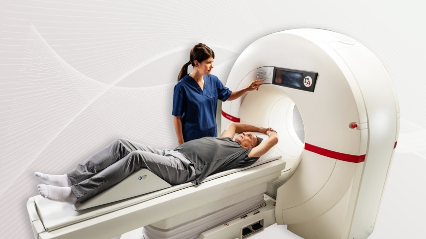
Cardiovascular health is paramount in maintaining overall well-being. As heart disease remains one of the leading causes of death worldwide, the importance of advanced imaging techniques cannot be overstated. Enter the cardiovascular CT scanner, a revolutionary tool in the early detection and management of heart conditions.
What is a Cardiovascular CT Scanner?
Definition and Overview
A cardiovascular CT (computed tomography) scanner is a specialized medical imaging device designed to capture detailed images of the heart and its blood vessels. Utilizing X-ray technology and advanced computer algorithms, these scanners produce high-resolution cross-sectional images, allowing for comprehensive evaluation of cardiovascular health.
How It Works
The scanner rotates around the patient, taking multiple X-ray measurements from different angles. These measurements are then processed by a computer to create detailed, three-dimensional images of the heart and blood vessels, providing insights into structure and function that are crucial for diagnosis and treatment planning.
Evolution of CT Scanners
Early Developments
CT scanners have come a long way since their inception in the 1970s. The first scanners were limited in speed and resolution, but they laid the groundwork for future innovations. Initially, CT imaging was primarily used for brain scans, but advancements soon expanded its applications.
Modern Advancements
Today, cardiovascular CT scanners are at the forefront of medical technology. Modern scanners boast faster imaging times, higher resolution, and greater accuracy. Innovations like multi-slice and dual-source CT have significantly enhanced their capabilities, making them indispensable in cardiology.
Components of a Cardiovascular CT Scanner
The Gantry
The gantry is the large, circular part of the scanner that houses the X-ray tube and detectors. It rotates around the patient to capture images from various angles.
X-Ray Tube
The X-ray tube emits X-rays that penetrate the body, varying in intensity based on tissue density. These variations are crucial for creating detailed images.
Detectors
Detectors capture the X-rays after they pass through the body, converting them into electronic signals that the computer system can process.
Computer Systems
Advanced computer systems compile the data from the detectors to construct detailed, high-resolution images of the cardiovascular system.
How a Cardiovascular CT Scan is Performed
Preparation for the Scan
Patients may be advised to avoid eating or drinking for a few hours before the scan. They should wear comfortable, loose-fitting clothing and remove any metal objects that could interfere with the imaging.
The Scanning Process
During the scan, patients lie on a table that slides into the gantry. They may be asked to hold their breath for short periods to minimize motion artifacts. The scanning process is quick, typically lasting just a few minutes.
Post-Scan Procedures
After the scan, patients can usually resume normal activities immediately. If a contrast dye was used, they may be advised to drink plenty of fluids to help flush it out of their system.
Benefits of Cardiovascular CT Scans
Non-Invasive Nature
One of the primary advantages of cardiovascular CT scans is that they are non-invasive. Unlike traditional angiography, which requires catheter insertion, CT scans do not penetrate the body, reducing the risk of complications.
High Accuracy and Detail
Cardiovascular CT scans provide high-resolution images that reveal even minute details of the heart and blood vessels. This accuracy is vital for diagnosing conditions like coronary artery disease.
Early Detection of Heart Diseases
By enabling early detection of heart diseases, cardiovascular CT scans allow for timely intervention, potentially preventing severe complications and improving patient outcomes.
Common Uses of Cardiovascular CT Scans
Coronary Artery Disease
Cardiovascular CT scans are particularly effective in diagnosing coronary artery disease by detecting blockages and narrowing of the coronary arteries.
Cardiac Structure Analysis
These scans can assess the structure and function of the heart, providing valuable information on conditions like congenital heart defects and cardiomyopathies.
Post-Surgical Assessment
After heart surgery, CT scans are used to monitor the success of the procedure and ensure there are no complications such as graft blockages or aneurysms.
Risks and Considerations
Radiation Exposure
While cardiovascular CT scans involve exposure to radiation, the levels are generally low and considered safe. However, repeated scans should be minimized to reduce cumulative exposure.
Contrast Dye Allergies
Some scans require a contrast dye to enhance image clarity. Although rare, allergic reactions to the dye can occur, so it’s important to inform the healthcare provider of any known allergies.
Claustrophobia and Anxiety
The enclosed space of the scanner can cause anxiety or claustrophobia in some patients. Open communication with the medical team can help manage these feelings.
Advances in Cardiovascular CT Technology
Multi-Slice CT Scanners
Multi-slice CT scanners can capture multiple slices of the heart in a single rotation, significantly reducing scan time and improving image resolution.
Dual-Source CT Scanners
Dual-source CT scanners use two X-ray sources simultaneously, allowing for faster imaging and better clarity, particularly in patients with high heart rates.
AI and Machine Learning Integration
Artificial intelligence and machine learning are increasingly being integrated into CT technology, enhancing image analysis, reducing errors, and providing more accurate diagnostics.
Comparing Cardiovascular CT with Other Imaging Modalities
CT vs. MRI
While both CT and MRI provide detailed images, CT is faster and better suited for detecting calcifications, whereas MRI excels in soft tissue characterization without radiation exposure.
CT vs. Echocardiogram
Echocardiograms use ultrasound to image the heart and are excellent for assessing heart function. However, they may not provide the detailed anatomical information that CT scans offer.
CT vs. Nuclear Imaging
Nuclear imaging involves the use of radioactive tracers to evaluate heart function and blood flow. It provides functional information that complements the structural detail seen in CT scans.
Preparation Tips for Patients
What to Wear
Patients should wear comfortable, loose-fitting clothes and avoid garments with metal zippers or buttons that could interfere with the imaging.
Eating and Drinking Guidelines
Typically, patients may need to fast for a few hours before the scan. Drinking plenty of water is recommended, especially if contrast dye will be used.
Medications and Allergies
Inform the medical team of any medications being taken and any known allergies, particularly to contrast dyes, to ensure proper precautions are taken.
Interpreting Cardiovascular CT Scan Results
Reading the Images
Radiologists and cardiologists analyze the images to identify abnormalities such as blockages, aneurysms, or structural defects.
Understanding the Report
The scan results are compiled into a report that outlines the findings and their implications. Patients should discuss this report with their doctor to understand the next steps.
Discussing Results with Your Doctor
A thorough discussion with the doctor helps clarify any questions and determine the best course of action based on the scan results.
Future Trends in Cardiovascular CT Scanning
Technological Innovations
Continuous advancements in CT technology promise even faster and more accurate imaging, with lower radiation doses and improved patient comfort.
Enhanced Diagnostic Capabilities
Future developments will likely enhance the ability to diagnose complex cardiovascular conditions with greater precision, potentially incorporating real-time imaging.
Personalized Medicine
As imaging technology evolves, it will play a crucial role in personalized medicine, allowing for tailored treatment plans based on detailed individual heart profiles.
Case Studies and Real-Life Applications
Success Stories
Numerous cases have demonstrated the life-saving potential of cardiovascular CT scans, such as early detection of critical blockages that were successfully treated.
Research Findings
Ongoing research continues to validate the effectiveness of CT imaging in diagnosing and managing heart conditions, leading to better patient outcomes.
Clinical Trials
Clinical trials are exploring new applications and technologies in cardiovascular CT scanning, promising further improvements and innovations in the field.
Conclusion
In summary, cardiovascular CT scanners represent a pinnacle of medical imaging technology, providing unparalleled detail and accuracy in diagnosing heart conditions. With continuous advancements, their role in cardiology will only grow, offering even more precise and personalized care.


